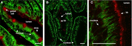
Figure 7.
Confocal images (63× objective) showing localization of Aqp1aa in middle (A) and posterior (B,C) segments of intestine from SW-acclimated Atlantic salmon. Sections were incubated with a cocktail of antibodies against Aqp1aa (red) and Na+, K+-ATPase (green). Lumen, goblet cells, nuclei, and brush border (bb) are indicated. Size bar = 20 μm.

