Figure 6.
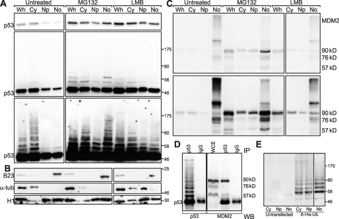
Subcellular fractionation of U2Os cells. (A) Western blot of fractions of U2Os cells treated as indicated at the top developed with anti-p53. (top) Camera image. (middle) Scaled camera image. (bottom) Film long exposure. (B) Fraction markers B23, α-tubulin (α-tub), and histone H1 are as indicated. (C) Western blot as in A developed with anti-MDM2. (top) Camera image. (bottom) Film image. (A–C) Black boxes are used to indicate that the data are derived from two separate membranes. (D) Immunoprecipitation (IP) of MG132-treated U2Os cells. Cell lysates were immunoprecipitated with either anti-p53 or purified murine IgG and probed on a Western blot (WB) with either anti-p53 or anti-MDM2. Black boxes are used to indicate that pieces of a single membrane were probed in parallel with the indicated antibodies. (E) Ubiquitylated p53. U2Os cells were untreated or transfected with 6×His-tagged ubiquitin (Ub) as indicated. Subcellular fractions were absorbed with Ni-NTA resin, and eluates were analyzed by Western blotting and detected with anti-p53. Black boxes are used to indicate that the lane for the nucleolar fraction (rightmost) was taken from a shorter exposure. Cy, cytoplasmic; Np, nucleoplasmic; No, nucleolar; Wh, whole fraction; WCE, whole-cell extract.

