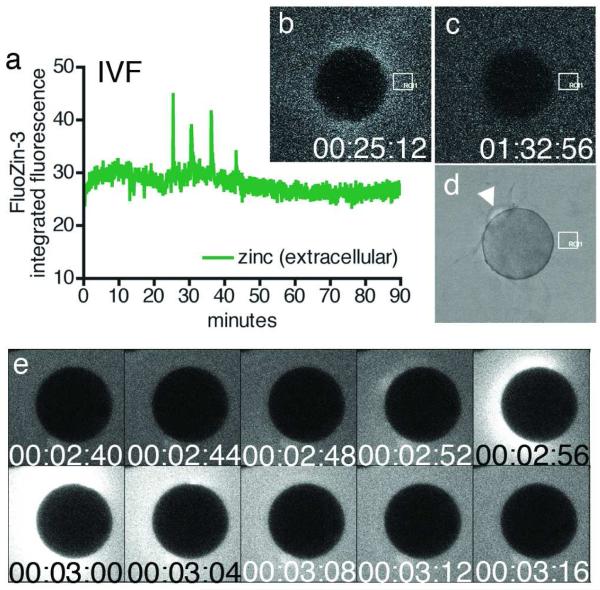
Figure 1. Zinc is released into the extracellular environment as an early fertilization event.
Changes in extracellular zinc concentration are readily monitored with FluoZin-3 during in vitro fertilization, and rapid, repetitive increases in fluorescence intensity were detected (a) by ROI analysis (denoted by white boxes in b-d). Each zinc spark can be distinguished in the time-lapse series (b, 00:25:12) against background fluorescence (representative example in c, 01:32:56). Successful fertilization was confirmed by the extrusion of a second polar body (d). Zinc sparks were also noted during strontium chloride-induced parthenogenesis (e, 00:02:56). Time is expressed as hh:mm:ss, wherein 00:00:00 represents the start of image acquisition.

