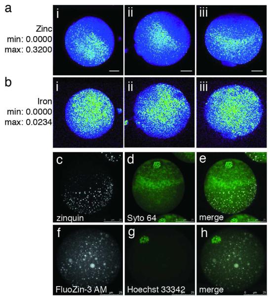
Figure 3. Zinc ions are cortically polarized in the mouse egg.
Total zinc, as detected by synchrotron-based x-ray fluorescence (a, b), is uniquely polarized in the unfertilized egg (a; i-iii represent replicates). This distribution is absent in the other essential transition elements, such as iron (b; i-iii represent replicates). The minimum and maximum range of each group of images are given units of μg/cm2. Labile (chelator-accessible) zinc, as detected by confocal microscopy (c-h), also has a hemispherical distribution in the live egg, as detected by two chemically distinct zinc fluorophores: zinquin ethyl ester (c-e) and FluoZin-3 AM (f-h). Co-staining with a DNA marker (Syto 64 in d, Hoechst 33342 in g) revealed that zinc was concentrated at the vegetal pole away from the meiotic spindle. The images depicted here are projected images of the complete Z-series; representative slices from these same Z-stacks confirming cortical localization of zinc are shown in Fig. S5. Scale bar = 20 μm (a, b) or 25 μm (c-h).

