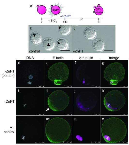
Figure 4. Sustained elevation of intracellular zinc availability following egg activation leads to reestablishment of metaphase arrest.
Eggs were activated with strontium chloride (SrCl2) then treated with zinc pyrithione (ZnPT) 1.5 hours later (a). At 6 h post-activation, control eggs form pronuclei (b, arrowheads) whereas the majority of eggs treated with ZnPT do not (c). When visualized by fluorescence, control eggs display decondensed DNA organized within a defined nucleus (d). F-actin is homogeneous around the egg’s cortex (e) and α-tubulin remains organized as a spindle midbody remnant (f), which is visible between the egg and the second polar body (g, merged image). In contrast, ZnPT-treated eggs display condensed chromosomes (h) adjacent to an area of concentrated, cortical F-actin (i). α-tubulin is organized in a metaphase-like configuration (j) around the chromosomes (k, merged image). This layout mirrors the subcellular arrangement in unfertilized eggs (l-o), which also display condensed chromosomes (l) overlaid with an actin cap (m), surrounded by a metaphase spindle (n). Scale bar = 80 μm (b, c) or 25 μm (d-o).

