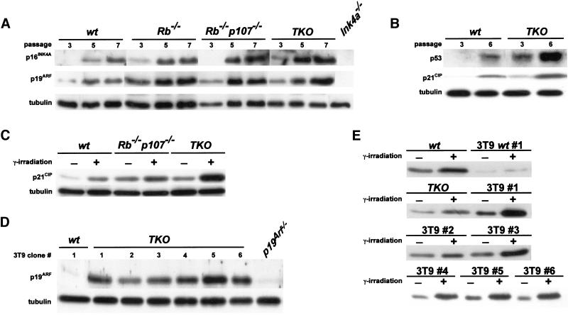
Figure 5.
Intact p19ARF/p53 pathway in immortal TKO MEFs. (A) Indicated MEFs were passaged according to a 3T9 protocol, and cell pellets were collected at passages 3, 5, and 7. Cell lysates were analyzed for p16INK4A, p19ARF and tubulin levels by immunoblotting. Ink4a−/− MEFs served as a negative control. (B) Expression of p53 and p21CIP in wt and TKO MEFs at passages 3 and 6. (C) Expression analysis of p21CIP in control and γ-irradiated early passage wt, Rb−/−p107−/− and TKO MEFs showing induction of p21Cip in all genotypes 24 h after irradiation. (D) Expression analysis of p19ARF in six independent TKO 3T9 clones (1–6). All clones expressed p19Arf, whereas a wild-type 3T9 clone (3T9wt, 1) had lost expression of this protein. p19Arf−/− MEFs served as a negative control. (E) Induction of p21Cip in γ-irradiated wt (1) and TKO (1–6) 3T9 MEF cell lines compared to γ-irradiated early passage wt and TKO MEFs.

