
Figure 3.
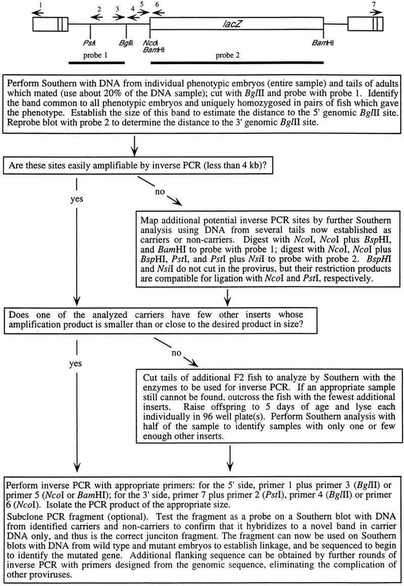
Isolation of genomic sequence flanking mutagenic insertions from multiple insert families. The schematic at top indicates the structure of the provirus along with the position of Southern blot probes and PCR primers.
Official websites use .gov
A
.gov website belongs to an official
government organization in the United States.
Secure .gov websites use HTTPS
A lock (
) or https:// means you've safely
connected to the .gov website. Share sensitive
information only on official, secure websites.
Isolation of genomic sequence flanking mutagenic insertions from multiple insert families. The schematic at top indicates the structure of the provirus along with the position of Southern blot probes and PCR primers.