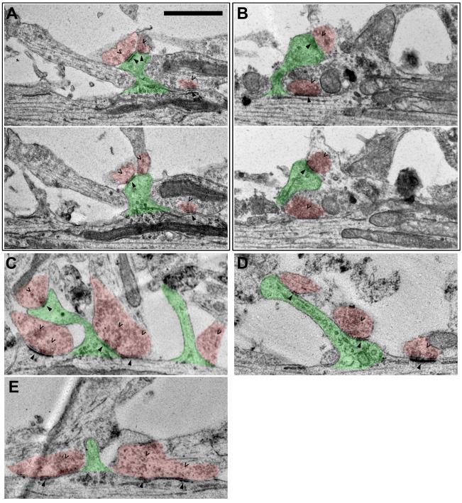
Figure 1. Serial section EM reveals dendritic spines protruding adjacent to shaft synapses.
Spines are shaded in green. Axo-shaft and axo-dendritic boutons are both shaded in red. Carets point to presynaptic vesicles in the boutons. Arrowheads point to postsynaptic densities in both dendritic shafts and spines. All neurons shown are at 3-4 weeks in vitro. Scale bar is 1 μm. (A, B) show two consecutive slices in the serial sections in which both pre- and postsynaptic features are visible. (C-E) show single slices of the serial sections in which both pre- and postsynaptic features are visible.

