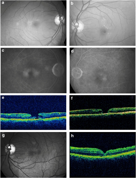
Figure 1.
(a and b) Fundus examination of both the eyes reveals a loss of foveal transparency and intra-retinal crystals suggestive of idiopathic macular telangiectasia type 2. The left eye also shows a FTMH, which was preceded by an inner lamellar defect. (c and d) Late-phase fluorescein angiogram shows typical parafoveal hyperfluorescence from the telangiectasia in both eyes. (e and f) Horizontal OCT scan of the left eye initially showed an lamellar macular hole, which dehisced into a FTMH after 14 months. (g and h) Fundus view and OCT scan at 11 months after surgery show complete closure of the macular hole without any residual neurosensory degeneration.

