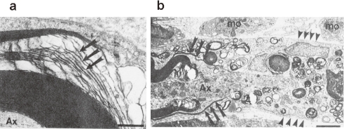
Figure 3.
Electron micrographs of rat sciatic nerve (a) 3 h and (b) 4 days after intraneural injection of anti-SGPG antibodies. (a) A large myelinated axon (Ax) with disintegrating leaflets of myelin (arrows), primarily along the intraperiod line. (b) An electron micrograph of the nodal region of an axon (Ax). To the left, normal appearing paranodal myelin is being stripped away by insinuating macrophage (mφ) processes (arrows) from a degenerating axon (Ax). Numerous macrophages are present in the endoneurial connective tissues. Scale bars: 0.5 µm (a), 2.5 µm (b).

