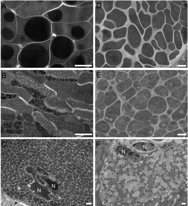
Figure 4.
Details from transmission electron microscopy of secretory vesicles from particular glands of T. regenti cercaria. A-C, "classical" method of sample treatment; D-F, high-pressure freezing/freeze substitution method. A+D, postacetabular vesicles. B+E, circumacetabular vesicles. C+F, vesicles in the head gland. N, nuclei of muscle cells surrounded by the head gland cell. Scale bars = 500 nm.

