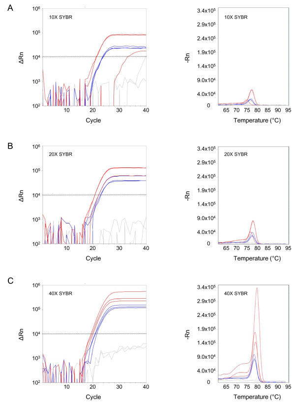
Figure 1.
Optimization of reagents for real-time PCR. Amplification curves (left panels) and melt curves (right panels) from Plasmodium falciparum gDNA spiked into uninfected blood and tested by real-time PCR. SYBR® Green dye concentrations of 10× (A), 20× (B), and 40× (C) were tested with 5% (red) and 10% (blue) volumes of blood in the PCR reaction. Uninfected blood serves as a negative control (gray). The stippled line marks the threshold for the amplification curves. Samples were run in triplicate.

