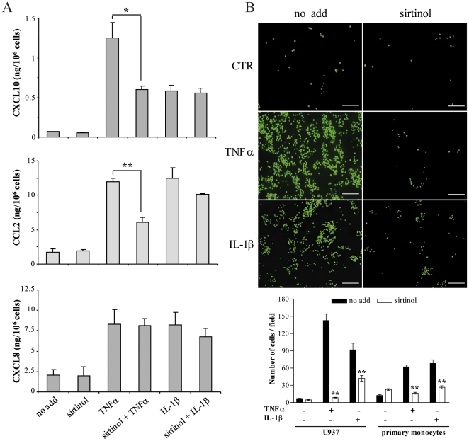
Figure 4. Chemokine secretion and monocyte adhesion in sirtinol-treated HDMEC.
(A) HDMEC conditioned medium was analyzed by ELISA after treatment with medium alone (no add) or sirtinol, followed by stimulation with the indicated cytokines. Results are the mean of at least three independent experiments and are given in ng/106 cells ± s.d; *p≤0.01, **p≤0.001, T-student test. (B) Fluorescence-labelled U937 cells or primary monocytes were plated on HDMEC pretreated or not (no add) with sirtinol, and exposed or not (CTR) to TNFα or IL-1β. Adherent cells were quantified as the mean number of fluorescent cells present in 10 randomly selected fields for each condition. Pictures of a representative experiment are shown in the left panels, bar = 50 µm. Data are expressed as the mean of three different experiments ± s.d (panel below), **p≤0.005, T-student test.

