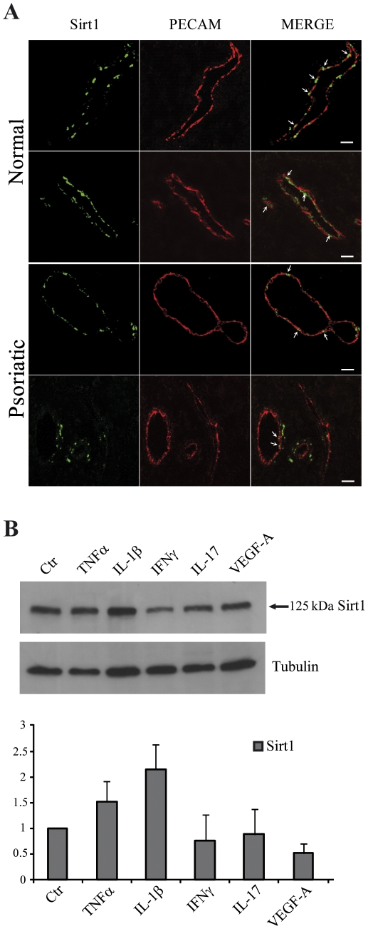
Figure 6. Sirt1 expression in skin microvessels.
(A) Sirt1 was stained with the primary antibody (left panels), and endothelial cells were put in evidence with a co-staining for the PECAM/CD31 transmembrane adhesion molecule (middle panels). Merged pictures are in the right panels. Each panel is a total projection of a confocal stack of images; bars = 20 µm. (B) HDMEC were treated with the indicated cytokines and the cell lysates were analyzed by immunoblotting with an anti-Sirt1 antibody. An antibody against tubulin was used as a loading control. A representative experiment is reported and the densitometric analysis of at least three different experiments is shown as the mean ± s.d.

