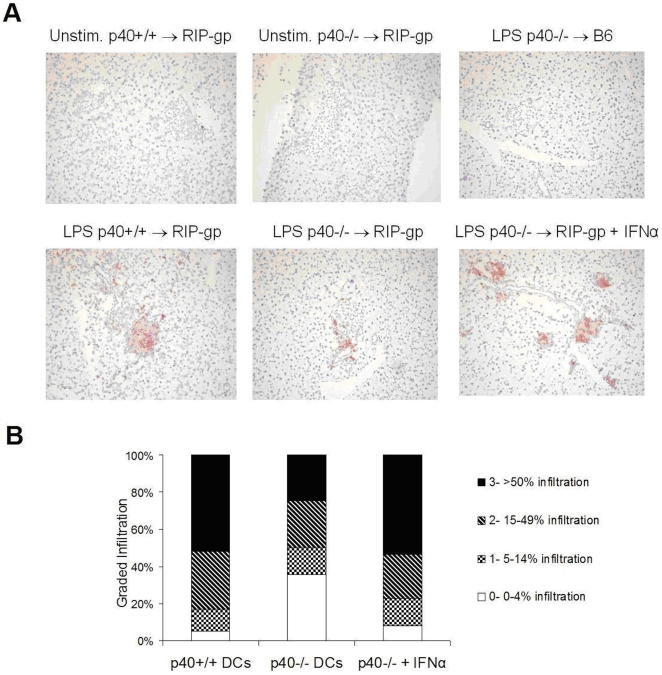
Figure 6. Exogenous IFNα enhances CD8 infiltration.
(A) Insulitis as assessed by immunohistochemistry of CD8 infiltrates (stained red) in pancreas sections five days after treatment. Representative sections from RIP-gp mice treated with p40+/+ (bottom left panel), p40−/− (bottom middle panel) peptide-pulsed BMDCs stimulated with LPS or p40−/− peptide-pulsed BMDCs stimulated with LPS with an additional i.v. injection of IFNα (bottom right panel) are displayed. For controls, representative sections of RIP-gp mice treated with unstimulated peptide-pulsed p40+/+ (top left panel) or p40−/− (top middle panel) BMDCs and C57BL/6 mice treated with LPS stimulated peptide-pulsed p40−/− BMDCs (top right panel) are shown. (B) Quantiation of CD8 infiltration, with the first 2 columns represented in figure 2 but reproduced here for comparison. Results are representative of a minimum of 5 mice per group, 100 islets per group from 2 independent experiments.

