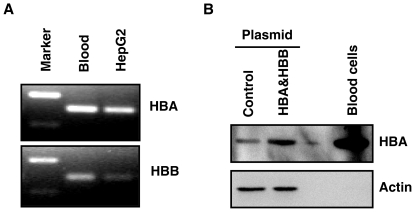
Figure 3. Expression of HBA1 and HBB in HepG2 cells.
A, HBA1 and HBB mRNA were detected by reverse transcription PCR. PCR products were separated on 2% agrose gel and visualized with ethidium bromide. PCR products were further confirmed by sequencing. B, HBA1 protein was detected by Western blot analysis. Whole cell lysates from HepG2 cells transfected with control or HBA&HBB expression plasmid were analyzed for HBA1 protein. Blood cells were used as the positive control. Beta actin was probed as a loading control.

