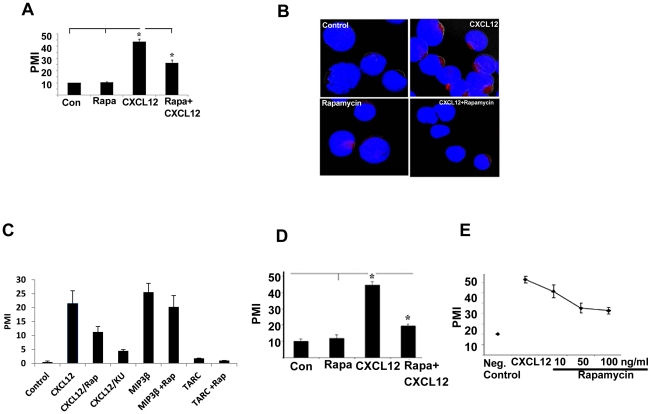
Figure 1. Rapamycin blocks CXCL12 induced migration and actin polymerization of T cells.
(A) Primary human T cells were labeled with calcein (5 µM) for 1 h in media and washed. Pretreatment of labeled cells was done with or without rapamaycin (100 ng/ml) for 1 h. Cells (1×10⁵ in 100 µl) were placed in the upper wells of 24-well transwell migration chambers with 5 µm pores (Corning, Corning, NY). In the lower wells, either medium alone or CXCL12 (100 ng/ml) was added to a total volume of 600 µl, and the chambers were incubated for 2 hours at 37°C in 5% CO2 incubator. Triplicate well determinations were performed for each treatment. The level of fluorescence of cells migrating across the chamber was assessed using a microfluorimeter. * indicates the value is statistically significant at p<0.05 level (n = 4). PMI indicates ‘percent migration index’. (B) Primary human T cells were treated with CXCL12 for 30 minutes in the presence or absence of pretreated rapamycin. Confocal microscopy was done according to the procedure mentioned in Materials and Methods to monitor actin polymerization. Red color indicates the actin polymerization. Representative images are shown from each treatment group. (C) Effect of rapamycin and KU-0063794 (KU) on CXCL12-induced cell migration, and effect of MIP3β and TARC in the presence or absence of rapamycin on the migration of resting T cells were performed as described in panel A. (D) Migration assay for CEM cells was performed similarly as primary T cells described above. (E) Dose response curve for rapamycin effect.

