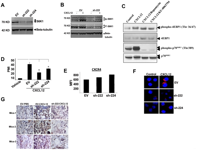
Figure 2. Role of mTOR in CXCL12-induced signaling and cell migration.
(A) Whole cell lysates from CEM stable clones carrying either empty vector (EV) or shRNA constructs (sh-222 and sh-224) were analyzed by western blot analysis to determine the levels of total p70S6K1. (B) Whole cell lysates from EV and sh-222 clones treated with CXCL12 for 30 minutes were analyzed by western blot analysis to determine the levels of phospho-p70S6K1. (C) CEM cells were pretreated with rapamycin (100 ng/ml) and KU-0063794 (1 µM) for 1 hour, and then CXCL12 treatment was done for 30 minutes. Equal amounts of whole cell lysates were analyzed by western blot analysis. (D) Migration assay for EV, sh-222 and sh-224 clones were performed similarly as primary T cells described in Fig. 1A. (E) Surface expression of CXCR4 for EV, sh-222, and sh-224 were determined by FACS analysis, and the levels are expressed as mean fluorescence intensity (MFI). (F) Confocal microscopy to monitor CXCL12-induced actin polymerization in EV, sh-222, and sh-224 clones were performed similarly as primary T cells described in Fig. 1B. (G) In vivo migration of EV and sh-222 clone mediated by CXCL12 was determined as described in Materials and Methods. Tissue staining from individual mice is shown here.

