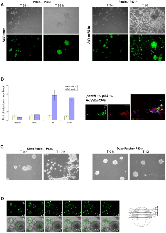
Figure 6. Neural differentiation of tumor spheres by miR-34a.
A. Differentiating effects of AdV-GFP-miR-34a on tumor spheres from Patch+/- P53+/- mice. Representative microscopy images and confocal GFP immunofluorescence staining of Patch+/- P53+/- mouse MB spheres at 24 h and 96 h from AdV-GFP-mock (left) or AdV-GFP-miR-34a (right) virus infections. B. Left: Real-time PCR analysis showing expression levels of the neural markers Nestin, MAP2, TUJ1, and GFAP in Patch+/- P53+/- tumor spheres at 96 h from infection with AdV-miR-34a or AdV-mock viruses. Fold changes are shown, calculated with respect to the gene expression of the AdV-mock infected tumor-spheres. Data are means ±standard deviations of 3 experiments, each carried out in triplicate. Real-time PCR reactions were normalized to β-Actin. Right: Representative immunofluorescence staining of Patch+/- P53+/- tumor spheres, differentiated following viral delivery of miR-34a, performed using an anti-GFAP antibody. GFP indicates the infection efficiency and the tumor sphere viability. C. Doxorubicin treatment of Patch+/- P53+/- and Patch+/- P53-/- tumor spheres. Representative microscopy images showing the neural differentiating phenotype observed only for the P53+/- tumor spheres. D. Left: Representative confocal GFP immunofluorescence staining of Patch+/- P53+/- tumor spheres at 24 h from AdV miR-34a virus infection. Arroweds denotes that the AdV-miR-34a virus efficiently infects only the cells located in the most external regions of the tumor spheres. Right: Illustration of the cell z-slices imaged.

