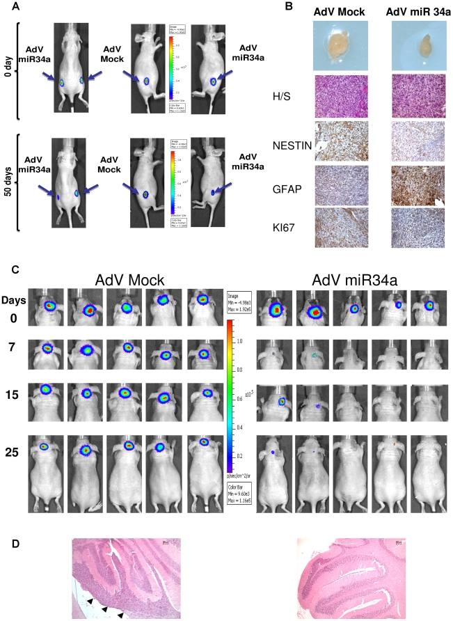
Figure 7. Orthotopic xenografts of MB Daoy cells overexpressing miR-34a by adenovirus infection: functional effects of miR-34a in vivo.
A. BLI of one selected mouse showing development of tumor burden over 50 days. Daoy-Luc cells previously infected with AdV-miR-34a or AdV-GFP-mock viruses were injected into the flanks of the nu/nu mice. B. Top to bottom: Tumor size, hematoxylin-eosin and immuno-histochemistry staining of Daoy tumors raised into the flanks of the nu/nu mice, for Nestin, GFAP and KI67. C. BLI of five mice injected in the fourth cerebellar ventricle with Daoy-Luc cells previously infected with AdV-miR-34a or AdV-GFP-mock viruses. Photon emission shows that within 25 days there is development and engraftment of the tumor burden with the AdV-GFP-mock that is greater than that with AdV-miR-34a. D. Hematoxylin-eosin staining of MB Daoy orthotopic xenografts raised in the nu/nu mice (left) and of a normal cerebellum (right). Arrowheads denotes tumor engrafment. Scale bar 100 µm.

