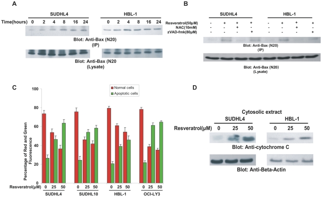
Figure 3. Resveratrol-induced mitochondrial signaling pathway in DLBCL cells.
(A) After treating with 50 µM Resveratrol for indicated time periods, HBL-1 and SUDHL4 cells were lysed and immuno-precipitated with anti-Bax 6A7 antibody for detection of conformationally changed Bax protein. In addition, the total cell lysates were immuno-blotted with specific anti-Bax polyclonal antibody. (B) HBL-1 and SUDHL4 cells were pre-treated with either, 10mM NAC and 80 µM z-VAD/fmk for 2 hours and subsequently treated with 50 µM Resveratrol for 8 hours. Cells were lysed and immunoprecipitated with anti-Bax 6A7 antibody and proteins were immunoblotted with Bax rabbit polyclonal antibody. (C) DLBCL cells were treated with and without 25 and 50 µM Resveratrol for 24 hours. Live cells with intact mitochondrial membrane potential and dead cells with lost mitochondrial membrane potential was measured by JC-1 staining and analyzed by flow cytometry as described in Materials and Methods. (D) SUDHL4 and HBL-1 cells were treated with 25 and 50 µM Resveratrol for 24 hours. Mitochondrial free cytosolic fractions were isolated and immunoblotted with antibody against cytochrome c and Beta-actin.

