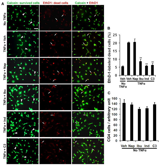
Fig. 1. RhoA-inhibiting NSAIDs dramatically protect differentiated CG4 cells from death induced by TNF-α.
A. The representative examples of differentiated CG4 cells were labeled with calcein (green, survival cell bodies and processes, arrowheads) and EthD1 (red, dead cell nuclei, arrows) from different groups treated with vehicle or drugs. In the absence of cytokine TNF-α, only a small number of dead cells were detected. Incubation with TNF-α for 8 hrs remarkably increased the number of dead cells in differentiated CG4 cells. However, treatments with Ibu, Ind or C3 transferase principally prevented cell death labeled by EthD1. In contrast, Nap did not have such a protective effect. Scale bar: 50 μm. B, Bar graph indicates percentage of dead CG4 cells labeled with EthD1 eight hours after TNF-α application. Treatments with Ibu, Ind or C3 transferase significantly protected CG4 cells from death induced due to TNF-α incubation. **P < 0.001, compared to the controls treated with vehicle or Nap (n=4 coverslips in each group). C, Graph shows effects of drug treatments for 9 hrs on the number of CG4 cells in the absence of TNF-α. Treatments with Ibu, Ind or C3 did not significantly alter the number of cultured CG4 cells.

