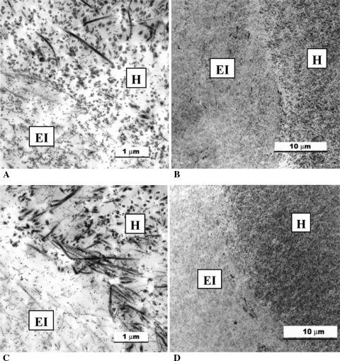
Fig. 6A–D.
Transmission electron microscopic images of the tissue-engineered implant and host cartilage interface at (A, B) 4 and (C, D) 8 weeks. The host cartilage was characterized by the presence of large collagen fibers, whereas the in vitro-formed cartilage had fewer collagen fibers that were smaller and thinner than those observed in the adjacent host tissue. At sites of integration, smaller fibers were seen admixed with the larger fibers. These interdigitating fibers were present by 4 weeks and seemed to increase in number with time. Both intact and necrotic chondrocytes were seen at the interface in some of the implants. EI = tissue-engineered implant tissue; H = interfacing host tissue.

