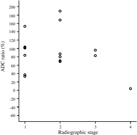
Fig. 3.
The graph shows diffusion changes in the epiphysis with radiographic stages of LCP. The dot plot shows that epiphyseal ADC remains increased at least until the healing stage in all cases (ADC - apparent diffusion coefficient; 1 - initial stage; 2 - fragmentation stage; 3 - healing stage; 4 - residual stage).

