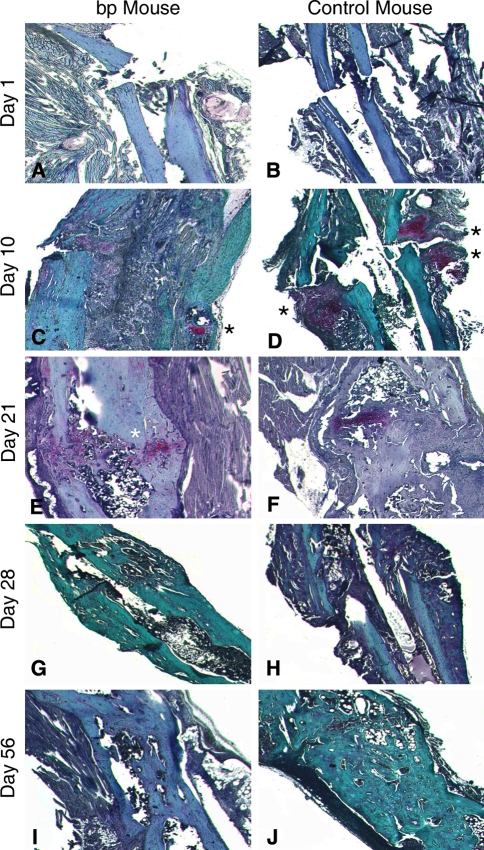
Fig. 5A–J.
Tibiae were isolated from bp and control wild-type mice, embedded, sectioned, and stained with Safranin O and Fast Green at Day 1 for (A) bp and (B) control wild-type mice; Day 10 for (C) bp and (D) control wild-type mice; Day 21 for (E) bp and (F) control wild-type mice; Day 28 for (G) bp and (H) control wild-type mice; and Day 56 for (I) bp and (J) control wild-type mice of healing postfracture. Blue or green staining indicates bone, pink staining denotes cartilaginous tissue or sutures, and purple staining indicates granular tissue. To highlight the presence of cartilage, it has been marked with a black or white asterisk. The (A–B) initial morphologic features of the fractures were similar in bp and control animals, but by Day 10, there was an increase in cellularity in (C) bp mice as compared with (D) controls. The wild-type callus contains cartilaginous deposits, whereas the bp callus does not. At Day 21 postosteotomy, cartilage formation was evident in (E) bp mice, whereas the (F) control fractures were undergoing remodeling of the cartilaginous precursor into bone. At (G–H) Days 28 and (I–J) 56 postfracture, the fracture was calcified, but remodeling of the callus was incomplete (Stain, Safranin O and Fast Green counterstained, original magnification, ×10).

