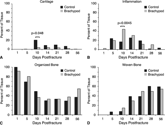
Fig. 6A–D.
The composition of the cartilaginous, inflammatory, and ossified callus was analyzed as a function of postfracture time in bp (gray) and control (black) treatment groups by morphometry. The temporal profile of cartilage deposition was similar in both treatment groups, peaking at (A) Day 10; however, substantially more cartilage was present in control calluses as compared with bp calluses at Days 10 through 21. The temporal profile of (B) inflammatory cell infiltration in the callus was similar in bp and control mice, peaking at Day 10 with greater amounts of inflammatory infiltration in bp fractures. There was a steep initial decrease in (C) organized bone in bp and control mice and increase in (D) woven bone after fracturing, with a plateau reached between Days 10 and 20 postfracture. Although the final amount of bone deposited was similar, there was more woven bone present in control animals at an earlier time as compared with bp mice.

