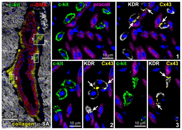
Fig. 2.
Vascular niches. Tangential section of a human epicardial coronary artery composed of a few layers of SMCs (α-SMA; red). At the interface between the adventitia and the SMC layer, three small clusters of c-kit-positive cells (green) are present and are shown at higher magnification in the adjacent panels. The c-kit-positive cells express KDR. Cx43 (arrows) is detected between c-kit-KDR-positive cells and fibroblasts (procoll; magenta).
(Taken from ref. 39 with permission)

