
Figure 1.
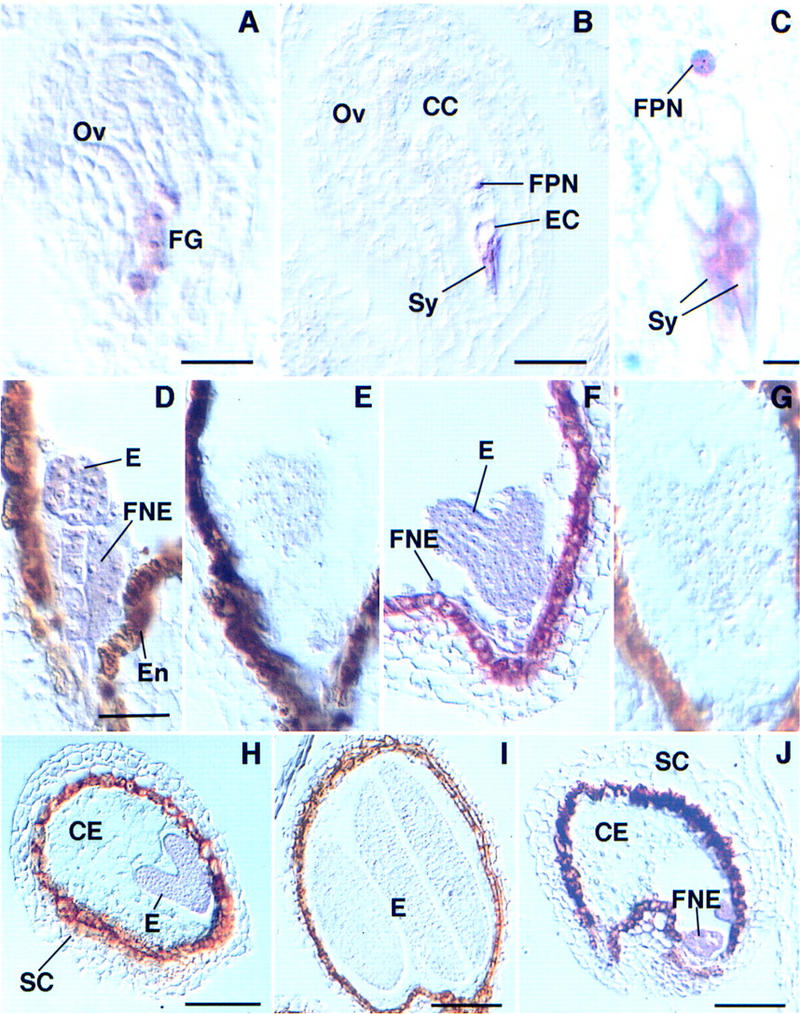
Localization of MEA mRNA in ovules and developing seeds of wild-type Arabidopsis. A-D, F, and H–J are hybridized with an anti-sense, E and G with a sense MEA probe. (A) Ovule (Ov) containing an eight-nucleated noncellularized female gametophyte (FG). The MEA transcript is present in the developing female gametophyte. (B) Mature ovule with unfertilized female gametophyte; MEA mRNA is present in the cytoplasm of the synergids (Sy), the egg cell (EC), and the central cell (CC) containing a homodiploid fused polar nucleus (FPN). (C) Detail of the synergids (Sy) and the fused polar nucleus (FPN) shown in B; the transcript is localized in the cytoplasm of the synergids, but appears closely associated with the fused polar nucleus. (D) MEA mRNA is localized in the globular embryo (E) and in the free nuclear endosperm (FNE) of a developing seed; artifactual staining in the endothelium (En) is seen in sense and anti-sense experiments. (E) Globular embryo hybridized with a sense MEA probe. (F) Heart-stage embryo hybridized with an anti-sense probe; the MEA transcript is localized in the embryo (E) and the free nuclear endosperm (FNE). (G) Heart-stage embryo hybridized with a sense MEA probe. (H) Seed showing an early torpedo embryo (E), the cellularized endosperm (CE), and the seed coat (SC); MEA mRNA is absent from the cellularized endosperm. (I) Seed containing a cotyledonary embryo hybridized with an anti-sense MEA probe. (J) Developing seed showing free nuclear endosperm (FNE), cellularized endosperm (CE), and the seed coat (SC); MEA mRNA is localized in the free nuclear endosperm. Bar, 17 μm in A; 22 μm in B; 4.3 μm in C; 35 μm in D–G; 51 μm in H–J.