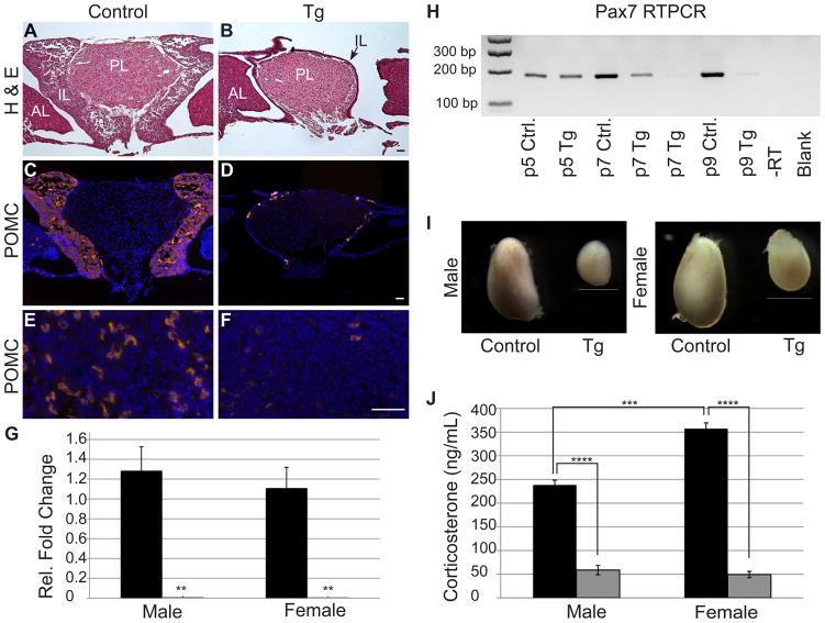
Figure 5.
Undifferentiated cells in Tg mice are incompatible with postnatal survival, resulting in adrenal dysfunction. Adolescent (p32) pituitaries were stained with hematoxylin and eosin to visualize morphology. Control (A) animals have a prominent IL, whereas Tg (B) animals have a very thin layer of IL cells (arrow) surrounding the PL. Nearly all of the IL lobe cells are immunoreactive for POMC (red) in the control (C) mice. In contrast, very few POMC-positive cells are observed in the IL of Tg mice (D). The same is true in the AL where POMC immunoreactive corticotropes are abundant in control mice (E) and nearly absent in Tg pituitaries (F). Similarly, qRT-PCR for Pomc reveals a significant decrease in Pomc mRNA in Tg adolescent pituitaries (gray bars) as compared to control littermates (black bars) in both males and females (G). Pax7 mRNA, which is exclusive to the IL in the pituitary, is present at p5 in both experimental groups and decreases dramatically by p9 in Tg pituitaries, indicating IL cells are no longer present at this age in Tg animals. Adrenals (I) of Tg males and females are reduced in size as compared to control mice. Stress-induced corticosterone levels (J) are significantly decreased in male and female Tg mice (gray bars) when compared to control mice (black bars). Photos taken at 100x (A–D) and 400x (E & F). Scale bars: 50μm (A–F) and 1mm (I). **: p<0.01. ***: p<0.001. ****: p<0.0001. n=3–5 (RT-PCR), n=5 (immunohistochemistry) and n=4 (corticosterone assay). PL=posterior lobe, AL=anterior lobe, IL=intermediate lobe.

