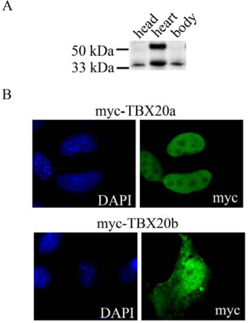
Figure 2. TBX20 isoforms show distinct tissue expression and subcellular localizations.
(A) Tissue from E12.5 ICR mice was dissected into head, heart, and body lysates. Lysates were analyzed by SDS-PAGE and western blot analysis. Antibody used was TBX20 (Sigma, HPA008192). TBX20a is 55 kDa. TBX20b is 33 kDa.
(B) COSM6 cells were plated on glass coverslips in a 24 well plate and transfected with 0.8 ug of DNA using Lipofectamine 2000 according to manufacturer’s protocol (Invitrogen). TBX20a and TBX20b were cloned into the pCMV-Tag3 construct to create a myc-tagged fusion protein (Stratagene). Cells were processed for immunocytochemistry using antibodies for myc (Cell Signaling). Coverslips were mounted onto glass microscope slides with DAPI counterstaining mounting solution (Vectashield).

