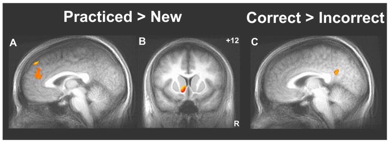
Figure 3.
Increased activity in targets of the mesolimbic dopamine pathway while performing the practiced sequence relative to the transfer conditions and in the posterior cingulate during periods of better performance. The Practiced-New contrast showed greater activity in the medial prefrontal cortex (A; whole-brain contrast, t > 4.5 and V > 327 mm3) and in the left ventral striatum (B; constrained search volume following ROI-AL of the striatum, t > 2.5 and V > 582 mm3; within the mask, left ventral striatum cluster volume is 731 mm3 and center of mass is at -8.4, +13.1, -1.5 mm). Activity in the posterior cingulate cortex (C) was higher during periods of more successful performance of the SISL task, i.e., was negatively correlated with number of errors (whole-brain contrast, t > 4.5 and V > 327 mm3; posterior cingulate cluster volume is 1234 mm3 and center of mass is at +0.6, -49.7, +28.8 mm).

