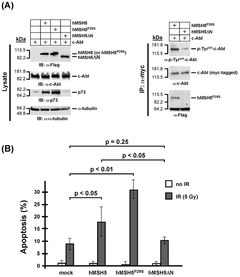
Figure 7.
The hMSH5-c-Abl interaction is important for stimulating c-Abl Tyr245 autophospho-rylation and IR-triggered apoptosis (A) Immunoblotting of the expression of various forms of hMSH5 proteins and their effects on p73 accumulation. 293T cells were transfected with c-Abl, or together with hMSH5, hMSH5P29S, or hMSH5ΔN. Cell extracts were prepared 48 h post-transfection, and levels of protein expression were analyzed by immunoblotting (left panel). The α-myc immunoprecipitates were used to analyze c-Abl autophosphorylation at Tyr245 and its interaction with hMSH5P29S or hMSH5ΔN (right panel). (B) TUNEL analysis of the effect of hMSH5ΔN on IR-triggered apoptosis. 293T cells were transiently transfected to express hMSH5, hMSH5P29S, or hMSH5ΔN 48 h prior to 5 Gy IR exposures, in which mock-transfected cells were used as controls. The rates of apoptosis were determined 24 h post-IR treatment. Mean percentages and standard deviations (error bars) were shown on the basis of three independent data points. P-values are indicated for statistical analysis.

