Figure 8.
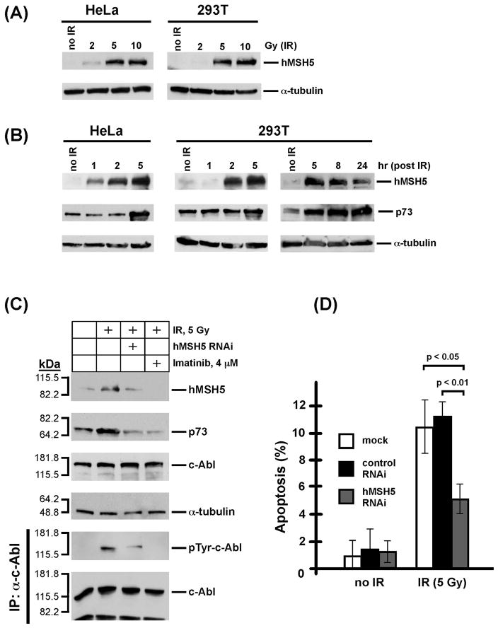
IR-triggered endogenous hMSH5 accumulation and apoptosis. (A) IR dose-dependent hMSH5 induction in HeLa and 293T cells. The levels of hMSH5 induction were evaluated 2 h after each treatment in reference to the level of α-tubulin. (B) Analysis of hMSH5 induction and p73 accumulation at various times after IR exposure. HeLa and 293T cells were irradiated with 5 Gy IR and cell lysates were prepared at various times as indicated. Immunoblot of α-tubulin was used as a loading control. (C) Effects of hMSH5 RNAi and imatinib on IR-triggered hMSH5 induction and p73 accumulation. 293T cells were transfected with hMSH5 sh-2 and treated with 5 Gy IR at 48 h post transfection, and the levels of proteins were evaluated 5 h after irradiation. Imatinib was used to inhibit c-Abl kinase activity. kDa, molecular weight (Mr) in thousands. (D) Preventing IR-triggered hMSH5 induction by RNAi compromised apoptosis. 293T cells were transfected with hMSH5 sh-2 or a control RNAi construct (pmH1P-Neo/NT, see Materials and Methods), of which the latter had no effect on hMSH5 expression (data not shown). Cells were exposed to 5 Gy IR at 48 h after transfection. Apoptosis was analyzed by TUNEL 24 h following irradiation. Error bars represent standard deviation from the mean of three data points. P-values are provided for significant differences.

