
Figure 6.
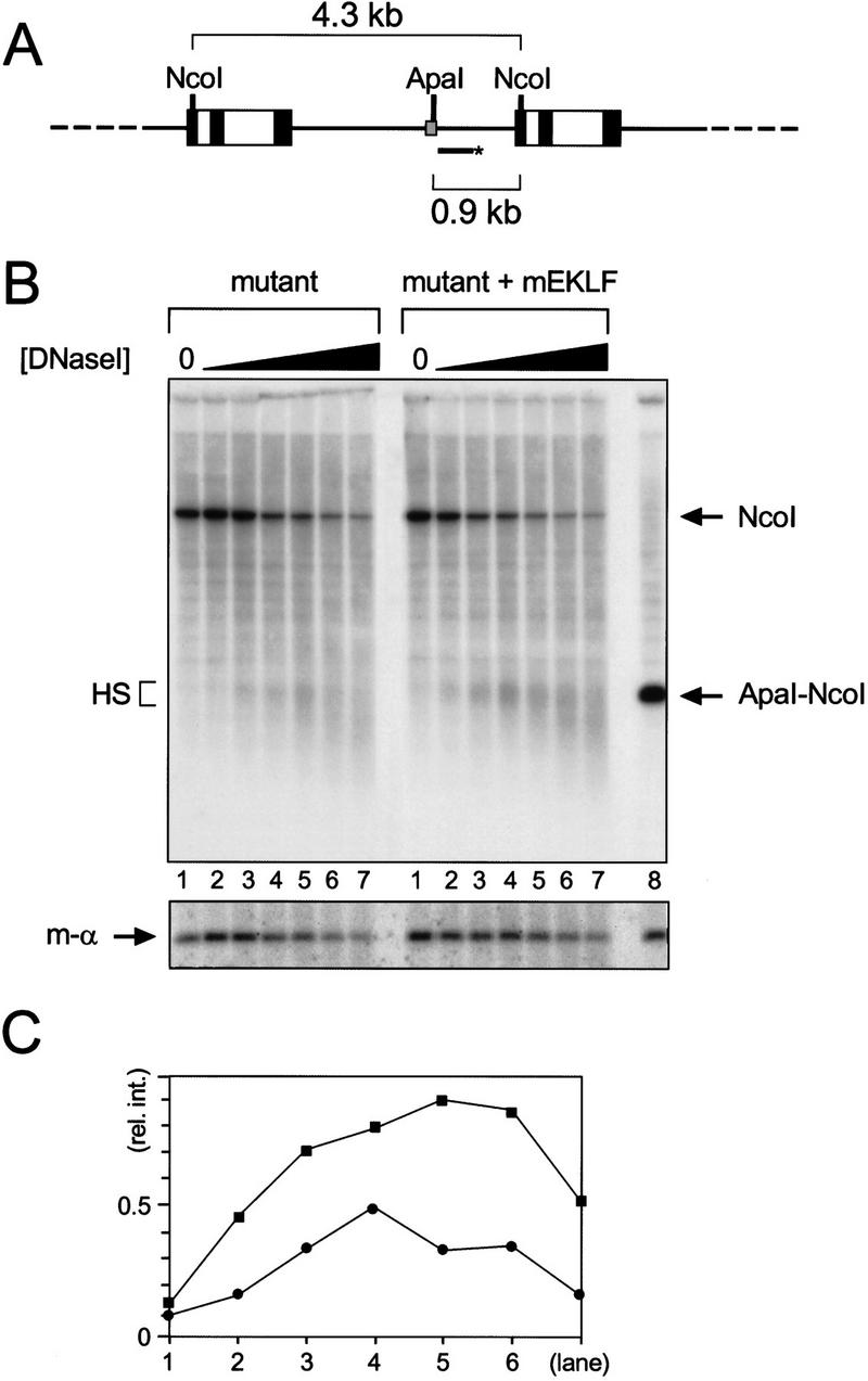
Hypersensitive site formation at the mutant fp 1–3 sequence in the presence of mEKLF. (A) Schematic drawing of the mutant reporter transgene array and the strategy used to detect hypersensitivity at fp 1–3. The HpaI–AccI probe does not detect the mEKLF–pEV transgene (Needham et al. 1992). The probe (bar with asterisk) hybridizes to the 4.3-kb NcoI repeat fragment of the mutant reporter gene. The 0.9-kb ApaI–NcoI fragment marks the position of the hypersensitive site (see B). (Other details are as for Fig. 1.) (B) Southern blot analysis of the hypersensitive site at the mutant fp 1–3 sequence. Nuclei were isolated from E13.5 fetal livers from mutant reporter and mutant reporter/mEKLF–pEV transgenics as indicated. Nuclei were digested with increasing amounts of DNase I, and DNA was purified, cut with NcoI, and analyzed by Southern blotting. The DNA in lane 8 was digested with NcoI and ApaI to mark the position of fp 1–3 (arrow, ApaI–NcoI). The repeat band of the mutant reporter transgene is indicated by an arrow (NcoI); the hypersensitive site at mutant fp 1–3 by a bracket (HS). (Bottom) The blot was stripped and rehybridized with a probe detecting an 0.85-kb NcoI fragment of the mouse α1-globin gene (m-α). (C) Quantitation of the hypersensitive site by PhosphorImager analysis. The signal in the hypersensitive site was divided by the signal in the corresponding mouse α1-globin band [(y-axis) relative intensity of the hypersensitive site] and plotted against the lane number (x-axis). (•) Mutant reporter; (█) mutant reporter plus mEKLF–pEV.