
Figure 3.
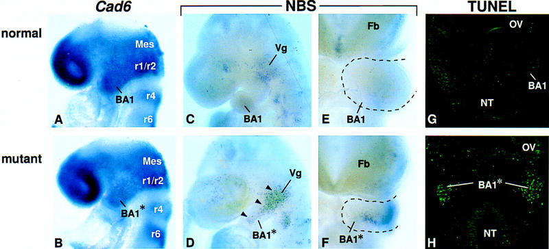
Analysis of neural crest cell migration and cell death in Fgf8;Nes–cre mutant embryos. (A,B) Expression of Cad6 at E8.5. (C–F) Cell death as assayed by Nile blue sulfate (NBS) staining in whole mount. Lateral (C,D) and frontal (E,F) views of E9.5 embryos. Black arrowheads point to areas of cell death in the mutant BA1. (G,H) Frontal sections of E9.5 embryos at the level of the maxillary primordium, stained for the presence of apoptotic cells using TUNEL. Abbreviations as in Figs. 1 and 2. (Mes) Mesencephalon; (NT) neural tube; (OV) optic vesicle; (r) rhombomere.