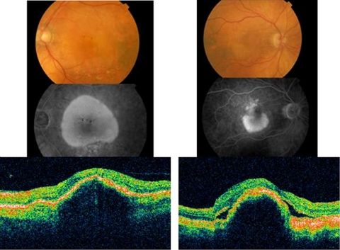
Figure 1.
A large serous PED is seen of the colour fundus photograph. There is increasing hyperfluorescence on fluorescein angiography, but no leakage. Blocked fluorescence corresponds to pigment seen on colour photography. A serous PED, without sub-retinal fluid or intra-retinal cysts, is seen on OCT examination. This lesion was felt to be avascular. A serous PED is seen below the fovea on colour and fluorescein angiography. There is speckled hyperfluorescence on angiography above the PED, consistent with occult CNV. There is sub-retinal and sub-RPE fluid on OCT examination. This lesion was felt to be vascularised.

