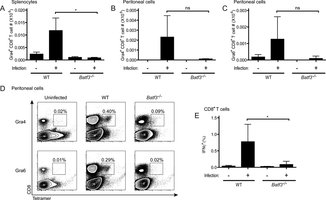
Figure 2. CD8+ T cell priming to T. gondii is defective in Batf3-deficient mice.
BALB/c wild-type and Batf3−/− mice were infected with T. gondii, sacrificed on day 8 after infection, and analyzed for CD8+ T cell priming by tetramer staining ex vivo (A–D) and intracellular cytokine staining following peptide re-stimulation in vitro (E). (A) Representative plots of Ld-GRA4 and Ld-GRA6 tetramer staining in the peritoneum, with percentage of total peritoneal cells that are tetramer positive shown. (B–D) Absolute numbers of CD8+ tetramer-positive cells in the peritoneum (B and C) or spleen (D) specific for GRA4 (B and D) or GRA6 (C) on day 8 after infection (n=3, representative of 2 independent experiments). (E) Absolute numbers of IFNγ-positive CD8+ T cells as measured by intracellular cytokine staining after overnight re-stimulation of whole splenocytes with the GRA4 peptide (n=5). (B–E) Data are represented as mean +/− standard deviation. Not significant (ns): P>0.05, *: 0.01<P<0.05.

