
Figure 1.
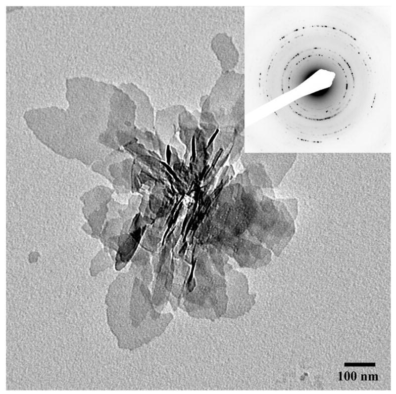
TEM micrograph and the corresponding diffraction pattern of randomly oriented plate-like apatitic crystals formed in the control experiment without proteins. The inset represents electron diffraction analysis. 86×86mm (300 × 300 DPI)
Official websites use .gov
A
.gov website belongs to an official
government organization in the United States.
Secure .gov websites use HTTPS
A lock (
) or https:// means you've safely
connected to the .gov website. Share sensitive
information only on official, secure websites.
TEM micrograph and the corresponding diffraction pattern of randomly oriented plate-like apatitic crystals formed in the control experiment without proteins. The inset represents electron diffraction analysis. 86×86mm (300 × 300 DPI)