
Figure 1.
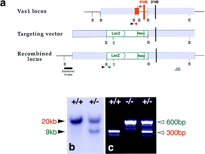
Disruption of the Vax1 gene by targeted recombination. (a) Maps of the wild-type Vax1 locus, the targeting vector, and the recombined allele. Relative positions of the exons coding for the two first helices (5′HB) and last helix (3′HB) of the homeobox are indicated by vertical bars. The map of the wild-type locus shows the deleted region in red, consisting of the start codon together with the exon coding for the first two helixes and amino-terminal part of the third helix of the homeobox and flanking intronic sequences. The map of the targeting vector shows the replacement of the deleted region by the β-galactosidase–neomycin cassette pGNA (green) (Le Mouellic et al. 1990). Arrowheads indicate the positions of the primers used for PCR genotyping. The dark line below the recombined locus denote the position of the genomic probe, external to the targeting vector, used to distinguish the EcoRV-generated 20- and 9-kb hybridizing bands on Southern gel of the wild type and recombined allele, respectively (b). Arrowheads in a denote the relative position of the primers used for PCR analysis and generating 300- and 600-bp amplification products for wild-type and recombined DNA, respectively (c). (E) EcoRV; (B) BamHI; (K) KpnI.