
Figure 5.
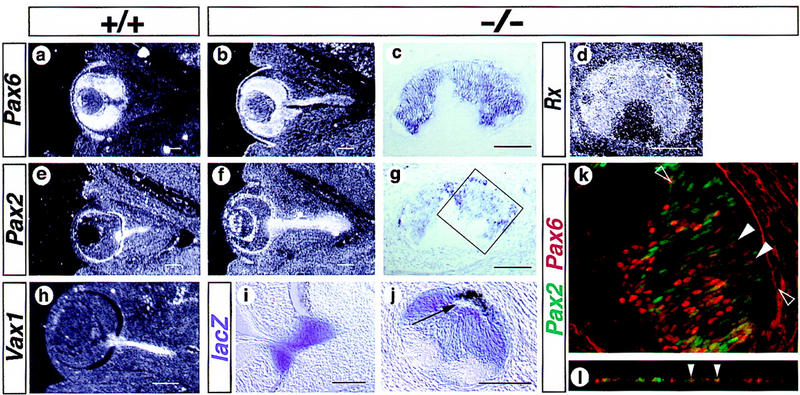
The Vax1 mutation affects the development of the optic nerve in homozygous mutants. (a–h) In situ hybridization with antisense probes as indicated on the figure, 13.5-dpc embryos; (i,j) β-galactosidase staining, 12.5-dpc embryos; (k,l) Pax2 (green) and Pax6 (red) immunocytochemistry, homozygous Vax1 mutant optic nerve at the temporal level, 13.5-dpc embryo; (l) confocal optical section through the z-axis of the section shown in k between the empty arrowheads. (c,d,g,j,k) Transverse sections of the mutant optic nerve. At 13.5 dpc, Pax6 expression is confined to the retina of the wild-type animal (a), whereas it remains expressed in the mutant optic nerve (b,c). Similarly, Rx is expressed ectopically in the mutant optic nerve (d). At 13.5 dpc, Pax2 expression is confined in the optic disk and optic nerve of wild-type animals (e) and remains expressed in these structures in Vax1 mutants (f,g). Similarly, Vax1 is expressed in the optic disk and optic nerve of wild-type animals (h) and the lacZ reporter gene is observed in these structures in Vax1 mutants (i,j). The expression of Pax2 and of lacZ indicates that the induction of the optic stalk occurred in Vax1 mutants. The thick epithelium observed ventrally (c,g,j) and the pigmented epithelium observed dorsally (j) in the mutant optic nerve therefore, could result from an abnormal development of the optic stalk rather than from an elongation of the retina. Pax2 and Pax6 are both expressed in the mutant optic nerve (k). However, protein expression is mostly exclusive at the cellular level. Coexpression is only detected in few cells expressing both proteins at low level (white arrowheads in k and l) suggesting a reciprocal inhibition of Pax6 and Pax2 and a participation of Vax1 in the down-regulation of Pax6. Dorsal is up in all pictures; rostral is left in c,d,g,j,k. The arrow in j points to the optic recess. Bar, 0.125 mm.