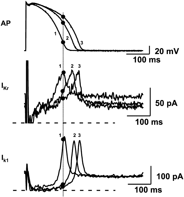
Figure 3.

When action potential duration has been prolonged previously by other means in dog papillary muscle (top panel), the positive feedback mechanism by which reduction of IKr (middle panel) and Ik1 (lower panel) further augments repolarization delay. Increased duration and thereby decreased rate of repolarization decreases the contribution of IKr and Ik1 currents to the repolarization process during the time course of the action potential, in turn producing a further slowing of repolarization. Numbers denote three recordings with different action potential durations. Dotted line identifies isochronal points on these recordings. IKr and Ik1 were measured from isolated ventricular myocytes subjected to action potential waveforms used as command signals in voltage-clamp conditions. AP, action potentials. Modified from Virág et al. (2009), with permission.
