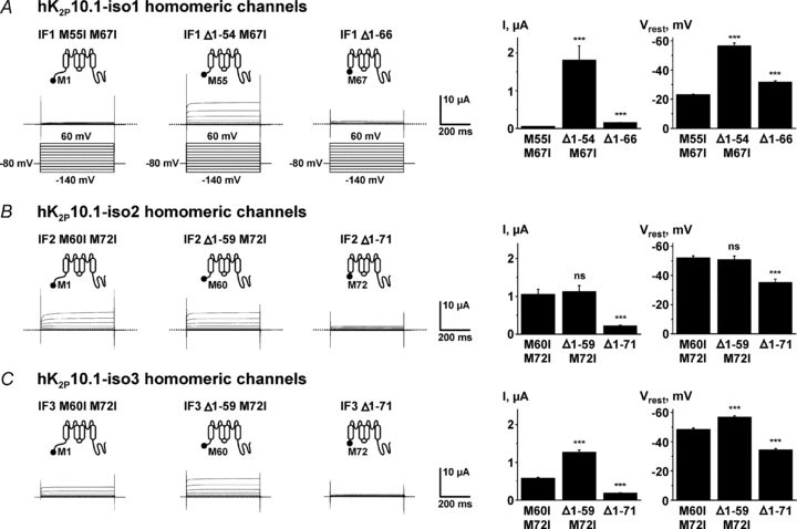
Figure 5. Biophysical characteristics of hK2P10.1 subunits studied in isolation.

Homomeric hK2P10.1-iso1 (A), hK2P10.1-iso2 (B), or hK2P10.1-iso3 (C) channels formed by indicated subunits were studied in Xenopus oocytes by two-electrode voltage clamp. Voltage protocols are depicted in panel A. Representative current families, mean outward currents at steady-state evoked by steps to 0 mV, and mean resting membrane potentials (Vrest) are displayed for groups of 16 cells. Error bars represent SEM; dotted lines indicate zero current levels. ***P < 0.001; ns, not significant versus respective full-length subunits.
