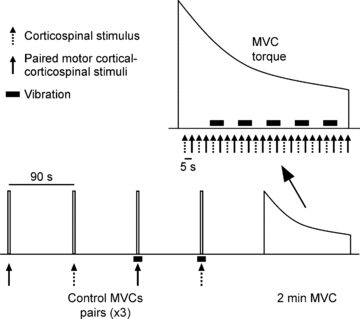
Figure 3. Schematic diagram of protocol A.

Twelve brief control MVCs were performed during which paired conditioning–test stimuli or a single test stimulus were delivered. Vibration (∼80 Hz) was applied to the distal biceps tendon on half of these efforts. Single and paired stimuli were delivered alternately at regular intervals whereas vibration was applied periodically during the sustained 2 min MVC. In protocol B, the test stimulus was a motor cortical rather than corticospinal stimulus.
