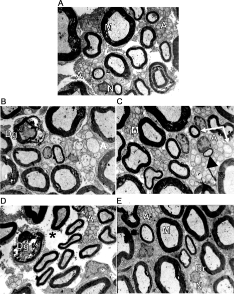
Figure 2. Electronic micrographics of sciatic nerve of rats who received chemotherapy with or without all-trans retinoic acid (ATRA) and controls.
(A) Myelinated axons with a myelin band according to their axonal diameter (M) in control group; preserved unmyelinated axons (AA) are shown. (B) Cisplatin group without ATRA treatment; degenerated myelinated axons (Dg) with altered myelin band and altered unmyelinated axons (A) were observed. (C) Cisplatin plus ATRA group showed normal appearance, myelinated axons with a myelin band according to their axonal diameter (M), conserved unmyelinated axons (A), and some had a diameter similar to those of small myelinated axons (arrows), probably indicating a remyelinated process, denoted by the thin myelin band (arrowhead). (D) In the paclitaxel-only treated group, degenerated myelinated axons (Dg) with altered myelin band and altered unmyelinated axons (A) are shown. There is endoneural damage denoted by spaces between myelinated axons and unmyelinated axon groups (asterisk). (E) Paclitaxel- and ATRA-treated group showed normal appearance, myelinated axons with a myelin band according to their axonal diameter (M), and conserved unmyelinated axons (A). There is a Schwann cell nucleus (N) surrounded by unmyelinated axon sprouts forming a regeneration band (Br). All electronic micrographics are at 6,860× and the quantifications are per square millimeter. Eight to 10 photographs were taken for each rat.

