
Figure 6.
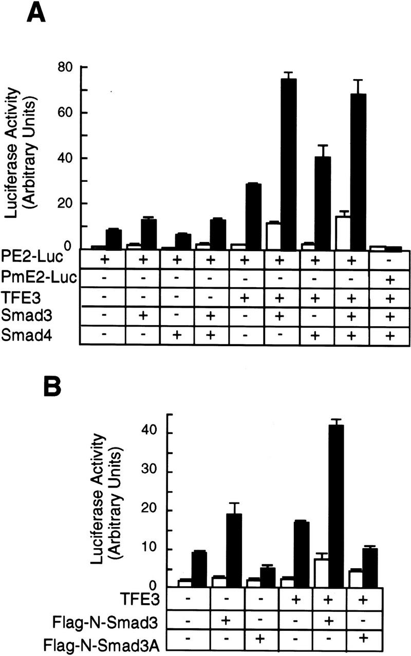
Synergy between TFE3 and Smad3 in the activation of the PE2 promoter. (A) Hep G2 cells were transfected as described in the legend to Fig. 5A. The following plasmids were used in transfection as indicated: 0.5 μg of plasmid encoding TFE3, 1 μg of plasmid encoding Smad3, and 1 μg of plasmid encoding Smad4; every well received 1 μg of PE2–Luc and 0.2 μg of pCMV–β-gal. The total amount of DNA was adjusted to 3.7 μg per well with pcDNA3. (B) Hep G2 cells were transfected with the following plasmids: 0.5 μg of TFE3, 1 μg of Flag-N–Smad3 or Flag-N–Smad3A; every well received 1 μg of PE2–Luc and 0.2 μg of pCMV-β. The total amount of DNA per well was adjusted to 3.7 μg with a control plasmid, pEXL–GFP. The cells were transfected, treated with (█) or without (□) TGF-β, and assayed as described for panel A.