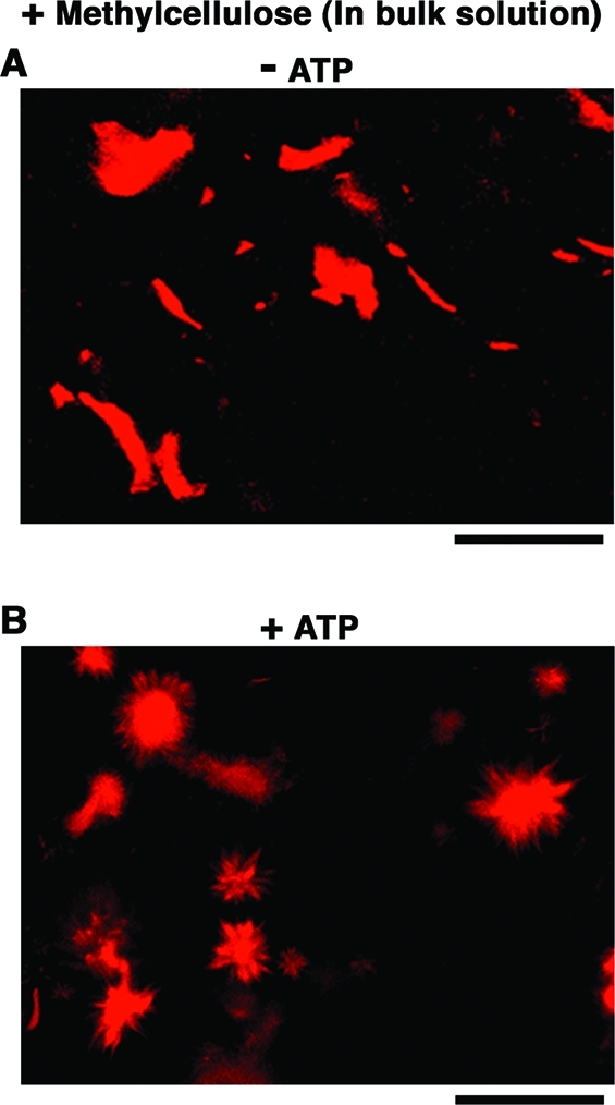
Figure 3.

Changes in the shape of actoHMM bundles induced by ATP in the bulk solution with 0.3% methylcellulose as observed by confocal fluorescence microscopy, where F-actin is labeled with rhodamine-phalloidin. (A) Without ATP, bundles have the typical structure. For a reference, length distribution of F-actin within actoHMM bundles is shown in Figure S4. (B) 30 min after the addition of 10 mM ATP, the formation of aster-like structures is noted. The transformation of actoHMM bundles in bulk solutions is not affected by the addition of α-hemolysin. Bars indicate 100 μm.
