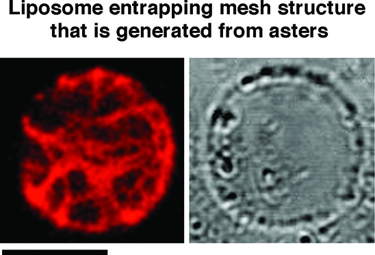
Figure 6.

Mesh structure inside cell-sized liposomes with actoHMM. The aster, which was formed in the bulk solution, was suspended and then entrapped in the giant liposome by the spontaneous transfer method (left, fluorescence; right, transmission). The lipid composition was DOPC/DPPC/cholesterol. The concentrations of F-actin and HMM are 10 and 20 μM, respectively. A 0.3% methylcellulose solution was used. Bar indicates 20 μm. Methylcellulose-formed actoHMM bundles are transformed to asters in the bulk solution by the addition of ATP. The asters are then encapsulated into cell-sized giant liposomes by the spontaneous transfer method using the pipetting procedure. By the suspension, the asters change in the mesh. The small spherical objects situated on the surfaces of liposomes in the transmission images are oil droplets in the water phase.(24) It should be noted that the encapsulated mesh is not transformed to an aster or situated on the inner surface periphery even after the addition of both α-hemolysin and ATP (Figure S3). The probability of the mesh generated inside was 75% among 100 liposomes.
