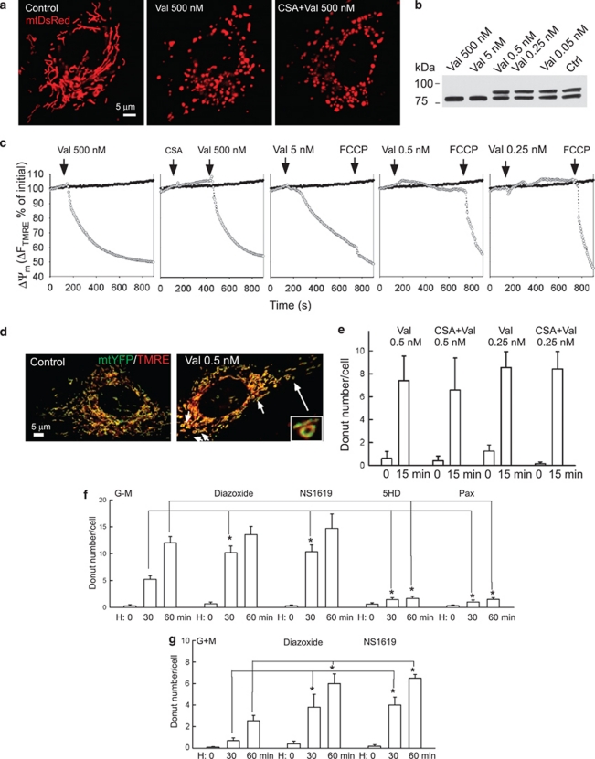
Figure 4.
Role of K+ channel activity in donut formation. (a) Images of mtDsRed-transfected H9c2 cells show that a high concentration valinomycin (Val) causes mitochondria swelling (middle) that is insensitive to CSA (right). (b) Effect of various doses of valinomycin on Opa1 cleavage (b) and on ΔΨm (c). Hollow circles show the H9c2 cells treated with valinomycin, whereas the filled symbols show the corresponding time control. (d) Images of mtYFP-expressing (green) and TMRE-loaded H9c2 cells show donut formation by a low concentration valinomycin (0.5 nM) that did not evoke ΔΨm loss, Opa1 cleavage and massive matrix swelling. (e) Lack of protection by CSA against valinomycin-induced donut formation. (f and g) Dependence of donut formation during H–R on K+ channel opening. The concentrations of drugs were as follows: diazoxide (KATP agonist, 100 μM), NS1619 (KCa agonist, 30 μM), 5HD (KATP inhibitor, 500 μM), paxilline (Pax, KCa inhibitor, 10 μM). H9c2 cells were pretreated with drugs for 20 min before hypoxia (H) (*P⩽0.004)

