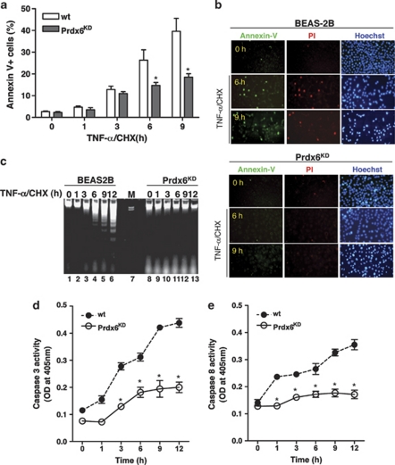
Figure 3.
Prdx6KD cells show strong resistance to TNF-α/CHX-induced apoptosis. (a) The wt BEAS-2B and Prdx6KD cells were treated with 100 ng/ml TNF-α/3 μ CHX for various times as indicated. The cells were stained for AnnexinV. The percentage of AnnexinV-positive cells was analyzed with the FACSCalibur system and determined with the CellQuest software. The results are expressed as mean±S.D. for triplicate assays. (b) The wt BEAS-2B and Prdx6KD cells were treated with TNF-α/CHX for 9 h. The cells were stained with AnnexinV/PI/Hoechst 33342, as described in Materials and Methods, and visualized with a fluorescence microscope. (c) The wt BEAS-2B and Prdx6KD cells were treated with TNF-α/CHX for various times as indicated. Cells were homogenized in 1 ml of lysis buffer. The genomic DNA extracts were prepared, as described in Materials and Methods, run on 1.8% agarose gels, and visualized under UV illumination. (d and e) The wt BEAS-2B and Prdx6KD cells were treated with TNF-α/CHX for different times as indicated. Caspase-3 (d) and caspase-8 (e) activities were measured using the CaspACE kit according to the manufacturer's instructions. The results are expressed as mean±S.D. for triplicate assays. P-values were calculated using t-test versus wt BEAS-2B (*P<0.05)

