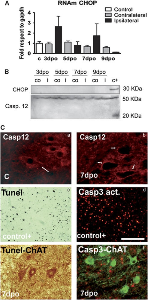
Figure 2.
(A) Histogram of mean values obtained by quantitative real-time PCR for Chop mRNA referred to Gapdh levels in the ventral horn of the spinal cord of control rats and in the ipsilateral and contralateral sides of root-avulsed rats. Samples were significantly different according to one-way ANOVA (*P<0.05), however, post-hoc analysis did not show up differences between ipsilateral and contralateral or control sides. (B) Western blot analysis of CHOP protein levels and full-length (50 kDa) or cleaved fragment (25 kDa) of caspase 12 at different time points in the ipsilateral (i) and contralateral (co) ventral horns of the spinal cord of root-avulsed animals. Note that we added a positive control from samples of animals submitted to spinal cord injury (c+).40 (C) Microphotographs of control and avulsed ventral horns immunostained for caspase 12 (a and b), and cleaved caspase 3 (red) (d and f) alone (d) or versus Choline acetyl transferase (ChAT, green) (f) at different times post-injury in control-positive animals (d) or root-avulsed ones (e and f). Note that caspase12 completely disappears from 7 dpo in the MNs and there is no colocalization between ChAT and cleaved caspase 3 immunostaining. Tunel staining (black) was also performed in a positive control section of brain tissue from an excitotoxic rat model52 (c), and in the root-avulsed model (e) colabeled for ChAT (brown). Bar=100 μm

